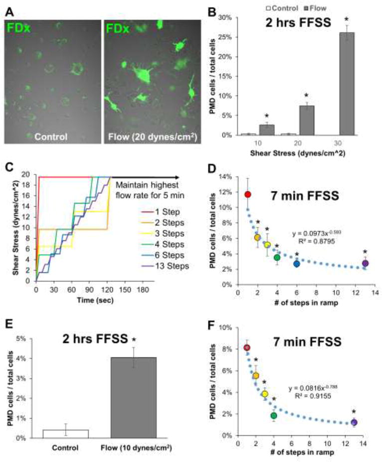
Figure 1. Osteocytes develop plasma membrane disruptions in vitro with fluid flow shear stress.

A–B) MLO-Y4 osteocytes were loaded with various magnitudes of fluid flow shear stress for 2 hours using medium containing 5% FBS, 5% BCS, 1% penicillin/streptomycin, and 1 mg/mL fluorescein-conjugated dextran. PMD-labeled osteocytes (green) were quantified from randomly collected confocal microscopy images (n=10 to 12 images per condition) after rinsing samples thoroughly in PBS to remove residual dye. *p≤0.05 vs. control for each experiment; each control/flow pairing is representative of three independent experiments. C–D) A 20 dynes/cm2 (2 Pa) shear stress was applied to MLO-Y4 osteocytes either immediately (1 step) or by gradually ramping up to this load (2 to 13 steps); PMD affected osteocytes were quantified as in panel B. *p≤0.05 vs. 1-step; representative of two independent experiments. E–F) Experiments in panels B and D were repeated with MLO-Y4 cells using medium containing 1% FBS, 1% FBS, 1% penicillin/streptomycin, 1 mg/mL fluorescein-conjugated dextran, and 1 mg/mL bovine serum albumin. *p≤0.05 vs. control or 1-step, representative of four independent experiments.
