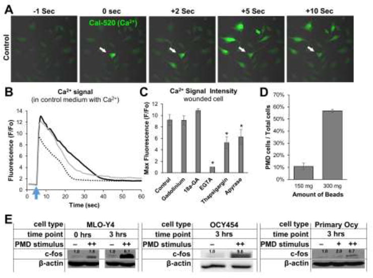
Figure 4. Plasma membrane disruptions initiate mechanotransduction in osteocytes.

A–B) MLO-Y4 osteocytes were loaded with Cal-520-AM Ca2+ dye and a 3 μm diameter PMD was created via laser as described in the Methods. Cells were imaged every second beginning 5 seconds prior to wounding and concluding 60 seconds after wounding. A) White arrow shows PMD site; other cells were not wounded. B) Ca2+ curves from 3 representative cells cultured in control medium are shown to demonstrate Ca2+ signaling magnitude and duration following a PMD; blue arrow indicates time of PMD formation. C) Quantification of Ca2+ signal intensity in wounded cells treated with various inhibitors, *p≤0.05 vs. control. D) Quantification of the number of cells wounded by the glass bead mechanical wounding protocol described in the Methods for either 150 mg or 300 mg of beads added to each dish; cells were quantified as in Figure 1B. E) Expression of c-fos in PMD-affected MLO-Y4 cells, OCY454 cells, or immortalized primary osteocytes; numbers above each band show expression normalized to actin +: 150 mg beads, ++: 300 mg beads. Each panel representative of n≥3 experiments.
