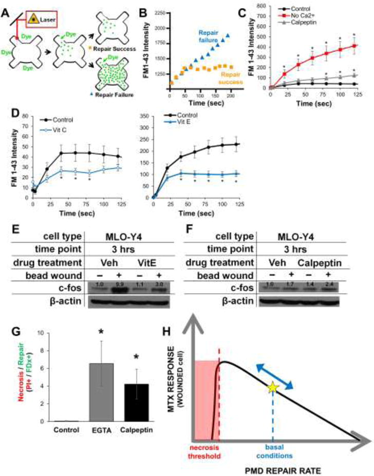
Figure 5. Osteocyte plasma membrane disruption repair rates can be enhanced or inhibited.

A–B) Schematic describing the laser-based wounding assay for analysis of PMD repair rate. Influx of FM1-43 dye near the membrane disruption is used to quantify repair rate; a fast plateau in fluorescence indicates rapid, successful repair, whereas increasing fluorescence over time indicates slow repair or repair failure. C–D) MLO-Y4 cell PMD repair rates were impaired by removal of extracellular calcium or following culture in calpeptin (which inhibits the membrane-bound calpain molecules involved in PMD repair), but were enhanced following culture in the antioxidants Vitamin C or Vitamin E. *p≤0.05 vs. time zero, n>20 cells measured for each condition. E–F) MLO-Y4 cells were cultured in repair enhancing (Vitamin E) or inhibiting (calpeptin) for 24 hours prior to wounding; expression of c-fos was conducted as in Figure 4E. G) MLO-Y4 cells were cultured in repair-inhibiting agents as described in the Methods and subjected to mechanical wounding by glass beads as described in Figure 4E. After wounding, cells were stained with propidium iodide to detect necrotic cells (i.e., unrepaired PMD) *p≤0.05 vs. control; 10 random images per condition were measured, and data are representative of three independent experiments. H) Proposed model for the influence of PMD repair rate on osteocyte mechanotransduction.
