
Figure 2.
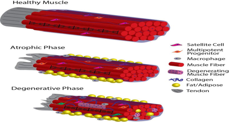
Schematic of progression of muscle pathology through different stages of RC disease. In healthy muscle with intact tendon (represented as a single fascicle connected to a tendon for simplicity), densely-packed muscle fibers are organized into fascicles by perimycium (translucent blue sheath), sarcomere length is maintained (black pattern on center-front fiber, not to scale), and muscle resident cells remain quiescent (note that cells are not to scale). With tendon disruption, RC muscle progresses to the atrophic disease stage, where muscle fibers become shorter, fiber cross-sectional area is reduced, and fat, fibrosis, and inflammation appear, while muscle architecture and overall muscle fiber and satellite cell numbers remain relatively constant. In the advanced, degenerative stage of RC disease, muscle fiber architecture is altered as sarcomeres remain chronically short, and damage and degeneration-regeneration becomes apparent (represented by the heterogeneous, hypercellular/myophagocytic light-pink fibers and centrally-nucleated, otherwise healthy fibers, respectively). The accumulation of inflammatory cells, collagen, and fat in both the inter-fascicular space (yellow spheres outside blue perimycium) and intra-fascicular space (yellow spheres between fibers) is more pronounced in the degenerative phase. Resident stem cell function is also disrupted in this stage of disease (represented in green in this schematic).