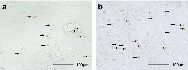
Fig. 1.
Histological sections (×400) of terminal deoxynucleotidyl transferase-mediated deoxyuridine triphosphate-biotin nick-end labeling (TUNEL) staining (a), and proliferating cell nuclear antigen (PCNA) staining (b) in ACL insertion after 2 weeks of knee immobilization. Brown cells (arrows) are TUNEL-positive chondrocytes and PCNA-positive chondrocytes. The TUNEL-positive and PCNA-positive rates were calculated by dividing positive cells by the total number of chondrocytes.

