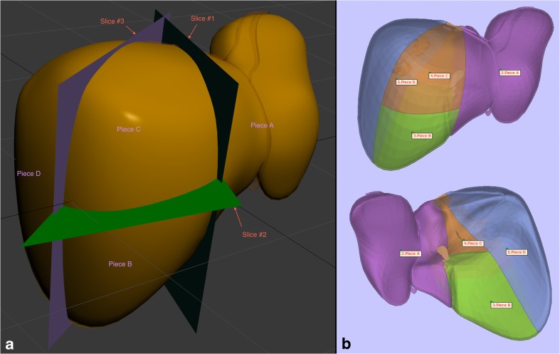
Fig. 1.
Liver surface. a Instructional figure for the graphic designer demonstrating the slice planes dividing the model into 4 pieces based on segmental anatomy. The reflection of the falciform ligament can also be seen, which, in part, demonstrates the delineation of the plane vertically dividing the left lobe of the liver. b Anterior and posterior views of the 4-piece model during quality control prior to 3D printing

