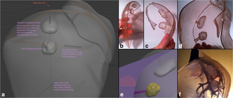
Fig. 4.
Liver abscess drainage and tumor biopsy tract. a Design instructions to the graphic artist in a view angle parallel to the plane separating the liver surface such that it is also along the tracts dividing them in half. b, c, d 3D-printed liver surface models that contain each half of the tracts, which when coming together, form a complete hollow tube. This affords the ability to easily place and remove a soft object in the site designated for the mass/tumor as well as fluid in the site for the abscess site. e Magnified view of the instructional figure for the graphic designer, showing cut plane passing through the middle of the hollow tubes connecting to the mass and abscess. f View of the final assembled 3D-printed liver model with its contents showing the lateral aspect of the right hepatic lobe with the abscess and mass representative sites seen through the transparent surface

