. 2017 Dec 31;19(4):284–290. doi: 10.7461/jcen.2017.19.4.284

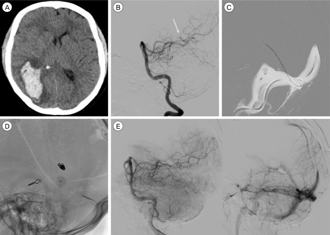
Fig. 1. A 50-year-old male with a right occipital hematoma. (A) Computed tomography scan showing the occipital hematoma. (B) Left vertebral artery angiogram demonstrates a small tentorial dural fistula supplied by the medial dural branch of the superior cerebellar to a deep cortical vein and ultimately draining into the straight sinus. Arrow shows drainage point in the straight sinus. (C) Trans venous navigation via the straight sinus into the draining cortical vein. (D) Lateral radiograph showing the final coil mass in the cortical draining vein close to the straight sinus. (E) Left vertebral angiogram demonstrates obliteration of the fistula with patent straight sinus.

Fig. 1