. 2017 Dec 31;19(4):284–290. doi: 10.7461/jcen.2017.19.4.284

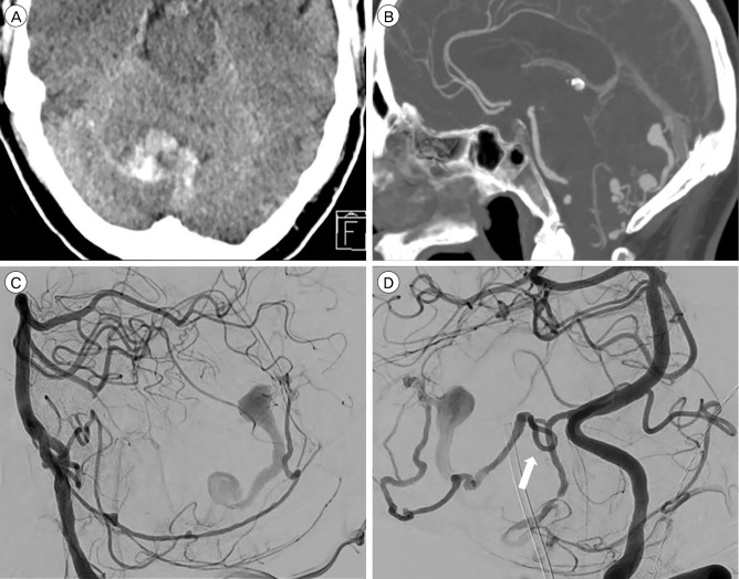
Fig. 3. A 63-year-old male presenting with right cerebellar hemorrhage. (A) Axial computed tomography (CT) scan demonstrating a right cerebellar hemorrhage. (B) CT angiography sagittal reconstruction demonstrating venous ectasia and fistulapoint at the torcula herophilii. (C) Left vertebral injection. Lateral angiogram demonstrating main arterial feeder from the posterior meningeal artery (PM). (D) Left vertebral injection. Arrow demonstrating PM loop where the PM is crossing the tentorial ridge. Distal endovascular access proved impossible because of this loop. Because no other viable arteries were available, the patient went for surgery.

Fig. 3