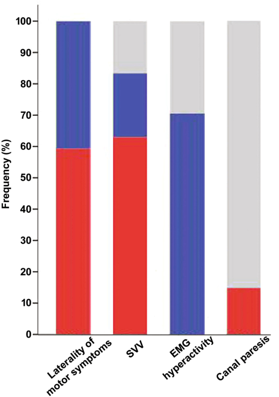
Figure 3.

Distribution of variables with directionality in PD-PS patients. Color bars represent the frequencies of PD-PS patients for each variable with directionality, including laterality of motor symptoms, subjective visual vertical (SVV), EMG hyperactivity, or canal paresis, respectively. Red color illustrates the frequency of PD-PS patients tilting to the less affected side, with ipsiversive SVV, ipsilateral EMG hyperactivity, or ipsilateral canal paresis. Blue color depicts the frequency of PD-PS patients tilting to the more affected side, with contraversive SVV, contralateral EMG hyperactivity, or contralateral canal paresis. Gray color represents the frequency of PD-PS patients who exhibit normal SVV, bilateral EMG hyperactivity, or no canal paresis.
