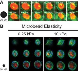
Figure 2.

Cell mimicking microparticles (CMMPs) within self‐assembled, stem cell spheroids can serve as force probes by monitoring their shape. (A): These three‐dimensional projection images from the Darling Lab at Brown University illustrate how highly compliant, 0.25 kPa CMMPs (red) deform in response to the contractile and adhesive forces of surrounding cells (green). Theoretically, an accurate reporting of the in situ stresses could be calculated based on the known mechanical properties of the CMMPs and their deformation from an original, spherical shape. (B): These two montages of confocal images (∼60 µm thickness, 7 µm steps) demonstrate that both 0.25 kPa (left) and 10 kPa (right) microbeads (red) are shuttled to the center of cell spheroids when coated in collagen. Cell nuclei (blue) and actin cytoskeletal structures (green) were stained with 4′,6‐diamidino‐2‐phenylindole (DAPI) and Alexa Fluor 488 phalloidin, respectively. Magnification: ×40.
