Figure 6.
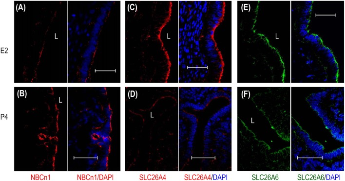
Effect of β-estradiol (E2) and progesterone (P4) on cellular expression of NBCn1, SLC26A4, and SLC26A6 in uteri of ovariectomized mice. (A,B) NBCn1 in the uterus from a mouse treated with E2 (A) or P4 (B). (C,D) SLC26A4 in the uterus treated with E2 (C), or P4 (D). (E,F) SLC26A6 in the uterus treated with E2 (E) or P4 (F). To compare the effects of E2 vs. P4 on the expression of a given transporter, immunofluorescence staining was always performed in parallel with the sections treated with either E2 or P4, and images were then acquired with the same set of parameters on the confocal microscopy. L: uterus lumen. Scale bars: 40 μm. Results are representative of 3–4 independent experiments with sections from three different mice for each treatment (E2 vs. P4).

