
Figure 3.
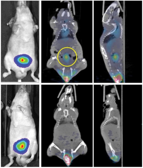
PET imaging of PS exposed in tumors.(p)(p)Notes: Mice implanted with tumors (PC3-luc) are injected with 124I-labeled 1N11 F(ab′)2 (PGN650) or control F(ab′)2 probes and imaged by bioluminescence in left panels and PET in middle and right panels. The yellow circle marks the tumor uptake of 124I-PGN650. Reproduced from Stafford JH, Hao G, Best AM, Sun X, Thorpe PE. Highly specific PET imaging of prostate tumors in mice with an iodine-124-labeled antibody fragment that targets phosphatidylserine. PLoS One. 2013;8(12):e84864.112(p)(p)Abbreviations: Ctrl, control; F(ab′)2, bivalent antigen-binding fragment of an antibody; I-124, iodine 124; luc, luciferase; PET, positron emission tomography; PS, phosphatidylserine.