Figure 5.
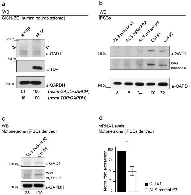
The dysregulation of Gad1 is conserved in human cell lines and iPSCs ALS patients derived. (a) Western blot analysis on human neuroblastoma (SK-N-BE) cell line probed for anti-GAD1, anti-GAPDH and anti-TDP in siTDP (TDP silenced) and siLuc (Luciferase ctrl). The same membrane was probe with the three antibodies and the bands of interest were cropped. Quantification of normalized protein amount was reported below each lane, n = 3. (b) Western blot analysis on human iPSCs probed for anti-GAD1 and anti-GAPDH in three clones derived from three different ALS patients (ALS patient #1 carrying the G287S mutation; ALS patient #2 carrying the G294V mutation; ALS patient #3 carrying the G378S mutation) and in two clones derived from two different healthy subjects (Ctrl #1 Ctrl #2). The same membrane was probe with the two antibodies and the bands of interest were cropped. In the two upper panels were reported two different exposition of anti-GAD1 signal. Quantification of normalized protein amount was reported below each lane. n = 3. (c) Western blot analysis probed for anti-GAD1 and anti-GAPDH on human differentiated motoneurons derived from iPSCs of an ALS patient (ALS patient #3 carrying the G378S mutation) and a healthy control (Ctrl #1 clone ND41864). The same membrane was probe with the three antibodies and the bands of interest were cropped. In the two upper panels were reported two different exposition of anti-GAD1 signal. Quantification of normalized protein amount was reported below each lane. (d) Real time PCR of Gad1 transcript levels normalized on Gapdh (housekeeping) in human differentiated motoneurons derived from iPSCs of an ALS patient (ALS patient #3 carrying the G378S mutation) and a healthy control (Ctrl #1 clone ND41864). n = 2, **p < 0.01 calculated by T-test. Error bars SEM.

