Figure 3.
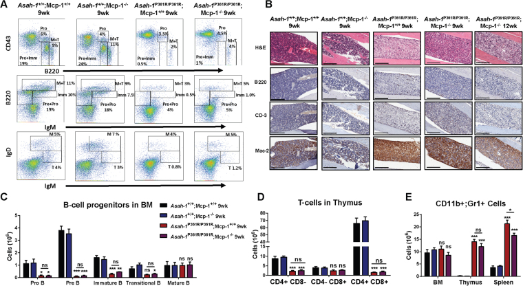
Perturbed hematopoiesis is retained in Asah1P361R/P361R;MCP-1−/− mice. (A) FACS plots showing B-cell lineage staining in BM from 9-week-old mice of all genotypes. (B) Histology staining of BM with H&E along with anti-B220, anti-CD-3, and anti-Mac-2 antibodies. (C) The absolute number of Pro-B, Pre-B, immature B, transitional B, and mature B cells in mice bone marrow (BM). (D) The absolute number of CD11b+ and Gr-1+ cells in mouse BM, thymus, and spleens. (E) The absolute number of CD4+ CD8−, CD4− CD8+, and CD4+ CD8+ cells in mouse thymus. 9- and 12-week-old mice were used for the histopathology analyses. Original magnification of BM micrographs at 10x where scale bar represents 300 µm. Spleen and thymus micrographs are at 4x magnification and the scale bar represents 100 µm. n = 3–4 mice at 9 weeks of age were used for FACS analysis and cell counts. ns (not significant), *p < 0.05 **p < 0.01, ***p < 0.001.

