Fig. 1.
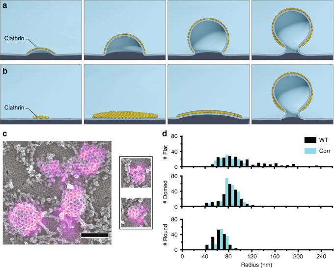
Mechanisms of membrane bending during CME and clathrin ultrastructure in unroofed SK-MEL-2 cells. a Schematic representation of CME in which membrane bending proceeds with a fixed radius of curvature during the addition of clathrin subunits. b Representation of CME in which clathrin first assembles into a flat sheet that remodels into a vesicle. c Correlative dSTORM and platinum-replica TEM images of fluorescently labeled clathrin (magenta) demonstrate a range of heterogeneous topographies even at the earliest stages of CME; scale bar is 200 nm. d The size distribution observed amongst clathrin structures in WT SK-MEL-2 cells (black) or SK-MEL-2 cells exogenously expressing clathrin light chain for correlative microscopy (blue). The inset shows ratios of average fluorescence associated with round vs. domed structures and domed vs. flat structures (SD shown for N = 3 cell membranes)

