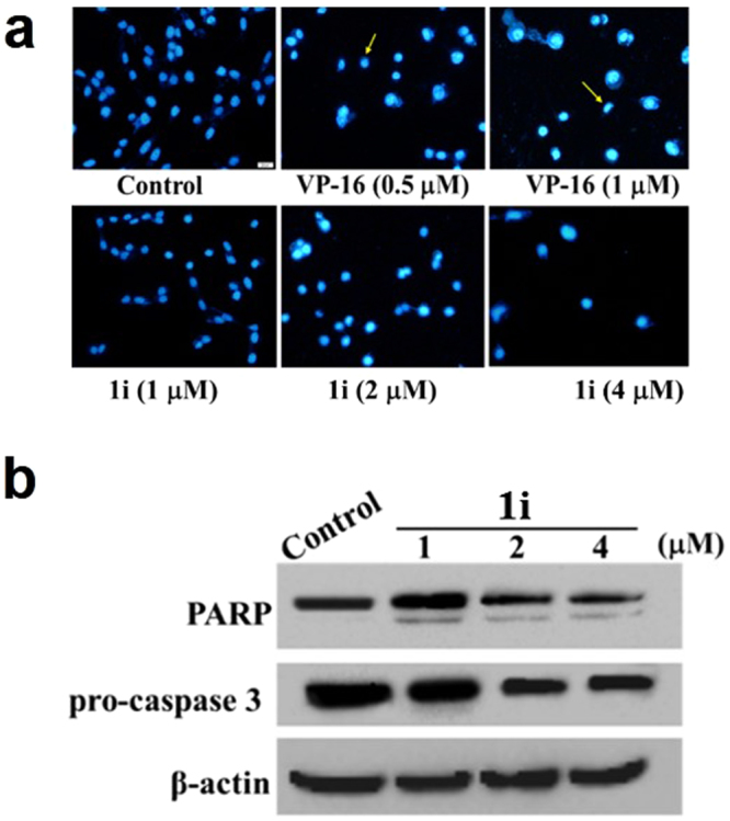
Figure 10.

Induction of apoptosis by active compound 1i and VP-16 at the indicated concentrations on PC-3 cells. (a) 72 h after the treatment of these compounds at the indicated concentrations, cells were fixed, washed with PBS, stained with Hoechst 33258, and analyzed for morphological characteristics associated with apoptosis by fluorescence microscopic analysis (×20). (b) Compound 1i triggered changes of pro-caspase 3 and PARP by Western blotting analysis. Cells were lysed after 72 h treatment with 1i at the concentration (1, 2 and 4 μM). The lysates were resolved on a 10% SDS-PAGE, transferred on to a nitrocellulose membrane and probed for cleaved caspase 3 and PARP. β-actin was used as a loading control.
