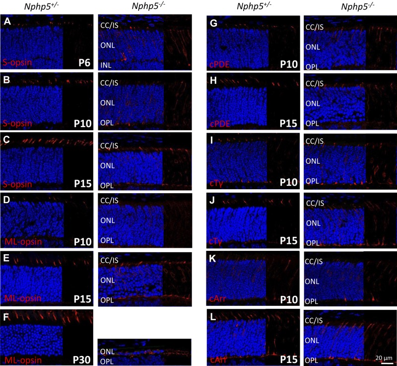
Figure 4.
Localization of cone OS proteins in Nphp5+/− and Nphp5−/− retina. A–C) Localization of S-opsin in heterozygous controls (left) and knockouts (right) at P6 (A), P10 (B), and P15 (C). D–F) Immunolocalization of ML-opsin in heterozygous controls (left) and knockouts (right) at P10 (D), P15 (E), and P30 (F). G–L) Immunolocalization of cone PDE6 (G, H), cone Tγ (I, J), and cone arrestin (K, L) in heterozygous (left) and homozygous knockouts (right) at P10 (G, I, K) and P15 (H, J, L). IS, inner segment; OPL, outer plexiform layer.

