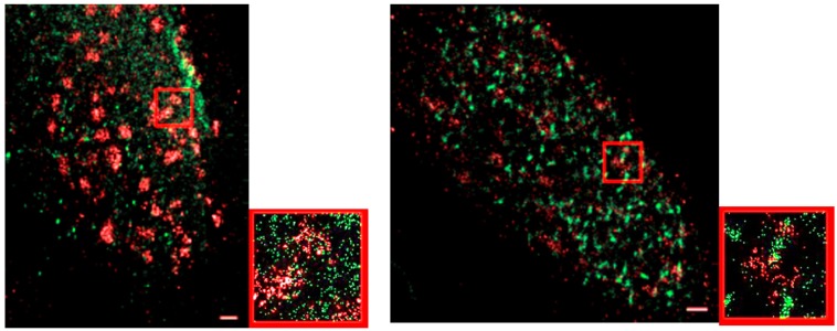
Figure 8.
Two color SMLM images of two MCF-7 cell nuclei after irradiation. Left images show the situation 30 min post irradiation; right images 180 min post irradiation. While γ-H2AX (red labelling) has already formed foci after 30 min, MRE11 (green labelling) is still more dispersed. After 180 min, both signals show strong clustering (foci formation). The small images are enlarged sections of the larger images (red rectangle).

