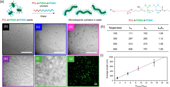
Figure 3.
(a) Schematic representation of epitaxial growth in water using PCL50-b-PMMA20-b-PDMA200 triblock copolymer. TEM micrographs of cylindrical micelles epitaxially grown from 40 nm seed micelles in water with a unimer/seed ratio of (b) 1, (c) 5, (d) 9, and (e) 15, using graphene oxide TEM grids.44 Scale bar = 1000 nm. (f) TEM micrograph (scale bar = 1000 nm) and (g) confocal microscopy image (scale bar = 20 μm) of fluorescently labeled cylindrical micelles epitaxially grown from seed micelles in water with a unimer/seed ratio of 15. Scale bar = 20 μm. (h) Length dispersity of cylindrical micelles. (i) Plot showing a linear epitaxial growth regime of cylinders with narrow length dispersities (error bars represent the standard deviation, σ, of the length distribution) in comparison to the theoretical length (dashed line).

