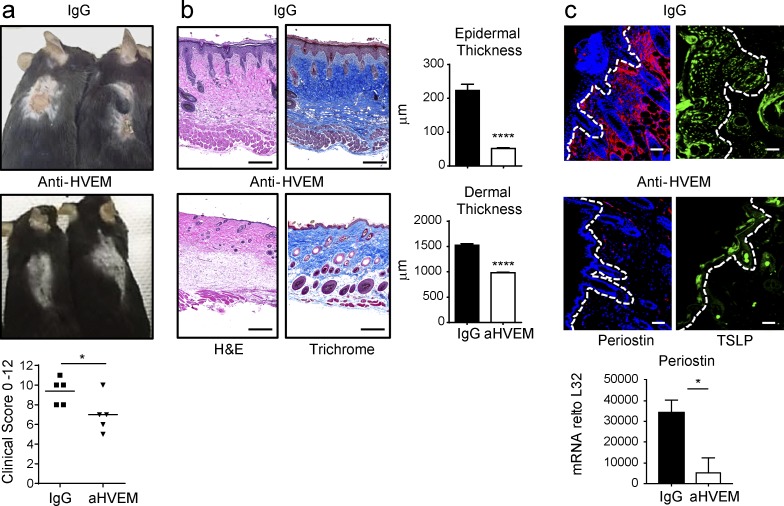
Figure 4.
Therapeutic blocking of LIGHT–HVEM reduces HDM-driven atopic dermatitis. (a–c) WT mice were sensitized epicutaneously with HDM/SEB and treated with anti-HVEM or IgG starting on day 7 and analyzed on day 14. (a) Clinical symptoms and scoring for skin eruption, scaling, bleeding, and redness from individual mice. (b) Skin sections stained with H&E or Masson’s trichrome blue and quantitated for epidermal and dermal thickness. Data represent means ± SEM from five mice. (c) IF staining of TSLP (green) and periostin (red). Dotted lines indicate epidermal-dermal border. mRNA expression of periostin in skin biopsy (means ± SEM). Data representative of 15 mice, with similar data in three experiments. Bars, 200 µm. *, P < 0.05; ****, P < 0.001. All statistical data were generated using the Mann–Whitney test.

