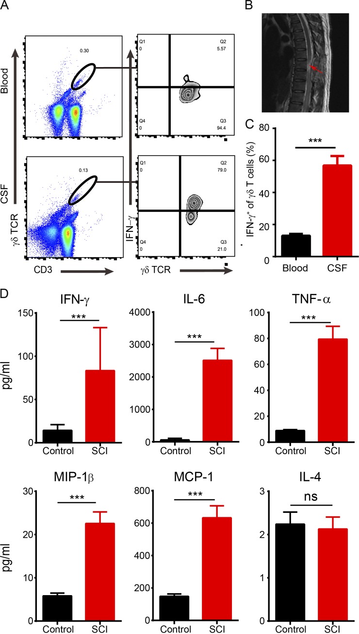
Figure 7.
Infiltration of γδ T cells and elevated levels of inflammatory cytokines in the CSF of SCI patients. (A) Using flow cytometry analysis, γδ T cells were enriched in the CSF and peripheral blood from SCI patients, and most of CSF-derived γδ T cells were positive for IFN-γ. (B) MRI image showing the injury site in a patient with SCI (arrow). (C) Statistical analysis showed a significantly higher ratio of IFN-γ+ γδ T cells in CSF than peripheral blood samples (P < 0.0001; n = 15). (D) The levels of IFN-γ, IL-6, TNF-α, MIP-1β, and MCP-1, but not IL-4, were significantly increased in the CSF from SCI patients compared with the control group (n = 20–22). Numbers represent frequencies (percentage) of cells in corresponding gates. Results (A and B) represent one of at least two repeated experiments. ***, P < 0.01. Mean ± SEM; Student's t test; ns, not significant.

