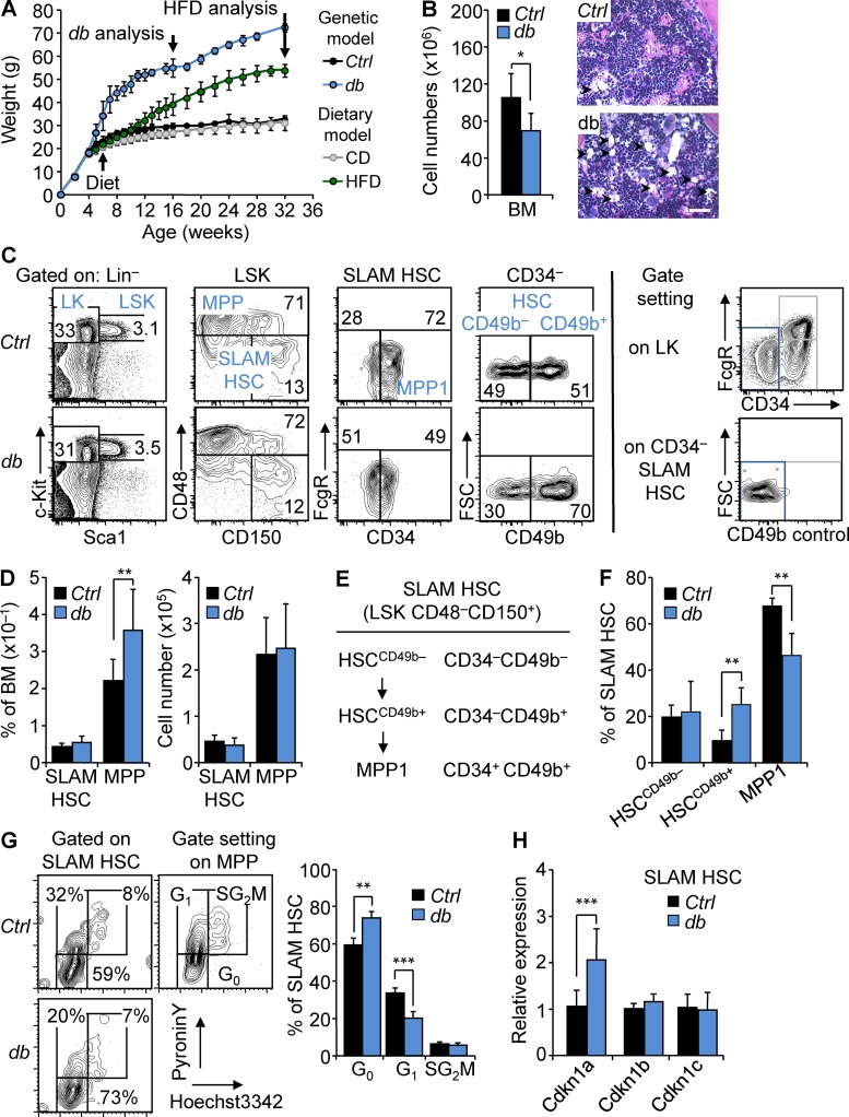
Figure 1.
HSCs maintain a highly quiescent state in obesity. (A) Kinetics of weight gain in genetic (db) and dietary (HFD) mouse models of obesity. db, n = 5; HFD, n = 8. (B) BM cellularity of 4-mo-old db mice compared with Ctrl littermates. n = 13/group. Images show H&E staining of the BM section from Ctrl and db mice (n = 3). Arrowheads indicate BM adipocytes. Bars, 100 µm. (C) FACS plots of HSPC populations in the BM of 4-mo-old Ctrl and db mice (n = 6). Right panels show strategies used to position CD34– and CD49b– gates. (D) Mean percentages (left) and absolute numbers (right) ± SD of HSPC populations in the BM of 4-mo-old Ctrl and db mice. n = 13/group. (E and F) Phenotypic definition and mean percentages ± SD of HSC subsets in the BM of 4-mo-old Ctrl and db mice. n = 6/group. (G) Representative FACS plots (left) and mean percentages ± SD (right) of Ctrl and db SLAM HSC distribution in cell cycle phases. n = 4/group. Two independent experiments. (H) qRT-PCR analyses for Cdkn1a, Cdkn1b, and Cdkn1c gene expression in SLAM HSCs isolated from 4-mo-old db mice. Results are expressed as fold change ± SD relative to Ctrl SLAM HSCs. n = 12 pools of 100 cells. Student’s t test; *, P ≤ 0.05; **, P ≤ 0.005; ***, P ≤ 0.0005. Two independent experiments.

