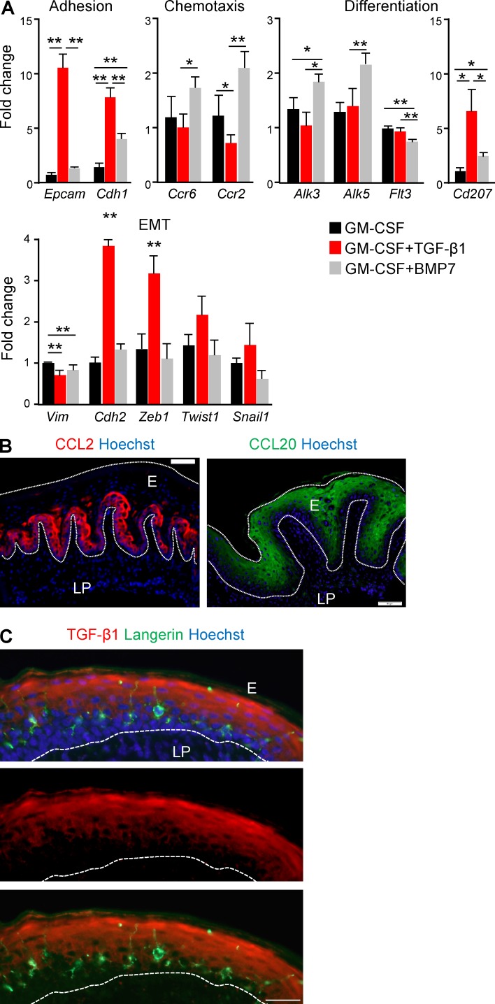
Figure 4.
Differential induction of genes involved in LC development by TGF-β1 and BMP7. (A) BM cells were incubated in serum-free SM in the presence of GM-CSF with or without TGF-β1 or BMP7. 5 d later, total RNA was prepared from the cultures and expression of the noted genes was quantified using quantitative RT-PCR. Bar graphs present the fold change in mRNA levels among the various treatments ± SEM (n = 5). Results of one experiments out of two independent experiments are presented. (B) Buccal cross sections prepared from 8-wk-old B6 mice were stained for CCL2 (red) or CCL20 (green) and Hoechst (blue). (C) Gingival cross section stained for TGF-β1 (red), langerin (green), and Hoechst (blue). Representative immunofluorescence image from one out of two-three independent experiments are presented. E, epithelium; LP, lamina propria. Dotted white lines indicate the basal and/or epical ends of the epithelium. Bars, 50 µm. *, P < 0.05; **, P < 0.001.

