Figure 7.
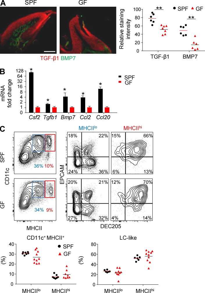
The impact of microbiota on mucosal LC differentiation is mediated locally rather than systemically. (A) Immunofluorescence confocal microscopy images of gingival cross sections from adult Swiss Webster SPF and GF mice stained for TGF-β1 (red), BMP7 (green), and Hoechst (blue). Graph indicates the relative staining intensity of the two cytokines using ImageJ software (n = 6–7). One representative experiment out of three independent experiments is shown. (B) Total RNA was extracted from gingival tissues of adult SPF and GF mice, and expression levels of the indicated genes were determined by quantitative RT-PCR. Bar graph presents the fold change in gene expression normalized to GF mice presented as the mean ± SEM. Data were pooled from three independent experiments, and each experiment included four separately analyzed mice. (C) Total BM cells from adult SPF or GF mice were incubated for 5 d in serum containing SM in the presence of GM-CSF and TGF-β1. Representative flow cytometry plots demonstrate the differentiation of CD11c-expressing MHCIIlo and MHCIIhi subsets as well as LC-like cells based on further expression of CD205 and EpCAM. Graphs present results pooled from three independent experiments shown as the mean values (n = 6 SPF and 9 GF mice). Bar, 50 µm. *, P < 0.05; **, P < 0.001.

