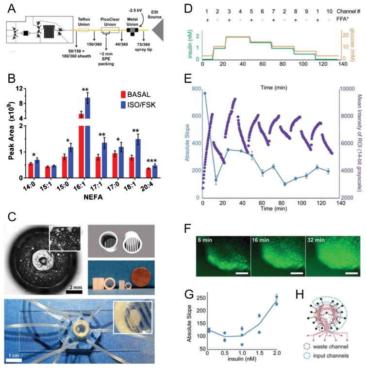
Fig. 1.
Recent examples of microfluidic systems for studying dynamic function of adipocytes and adipose tissue. (A) Schematic of microfluidic device for 3T3-L1 adipocyte stimulation and sampling, coupled to solid phase extraction and mass spectrometry (SPE-MS), for monitoring non-esterified fatty acid (NEFA) secretion profiles from adipocytes [49]. (B) Average NEFA secretion profiles during basal and isoproterenol/forskolin stimulation of adipocytes using the chip in (A) for stimulation and sampling. (C) On-chip culture systems for primary adipose tissue [42]. Primary mouse adipocytes were cultured in a 3D collagen matrix (top left) on an 8-channel microfluidic sampling device. Alternatively, explants were trapped using customized traps. 3D CAD rendering (top right) is shown with 3D-printed explant traps (middle right). (D) Temporal program of combined insulin, glucose, and fatty acid inputs to adipose explants using a microfluidic multiplexer (μMUX) device [39]. (E) Real-time fatty acid uptake responses from treatments in (D) showed insulin-dependent exchange rates. (F) Representative fluorescent images of on-chip explants used to generate data in (E). (G) Compiled data from (E) showed insulin-dependent fatty acid exchange dynamics. (H) Assignment map of the programmable, automated μMUX device, showing the 10 required input channels and one output waste channel used for (D)–(G). Reproduced from references 39, 42, and 49 with permission from the Royal Society of Chemistry and from Springer Publishing Company

