Figure 1.

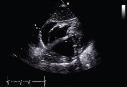
Initial echocardiographic examination – arrow points toward pericardial effusion 2 cm thick located next to the right atrium and atrioventricular groove

Initial echocardiographic examination – arrow points toward pericardial effusion 2 cm thick located next to the right atrium and atrioventricular groove