
Fig. 8.
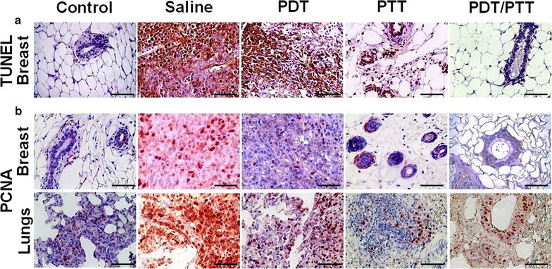
In situ apoptosis detection by TUNEL staining and immunolocalization of PCNA positive cells. The brown dye colored cells represent both TUNEL (control and tumoral breast tissues) and PCNA positive cells (breast and lung tissues). The sections were prepared 30 days from the last mice treatment with the phototherapies. a For TUNEL, only in breast tissue, these positive cells can be observed mainly in the saline group without an expressive labeling of cells for the combined PDT/PTT group. b Proliferating cell nuclear antigen labeled cells were natively found in lung tissue even in the control group. Breast tissue presented higher labeling in the saline group and lower in the treated groups, PDT and PTT only and combined PDT/PTT therapies, suggesting the potential of the therapies. Magnification ×400, scale bar = 100 µm. There was no statistical significance between the groups (p > 0.05)