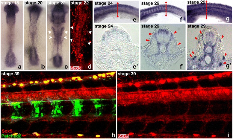
Fig. 4.

Expression of medaka sox5 during embryogenesis. a to c and e to g Medaka sox5 expression investigated by whole-mount in situ hybridization or d, h, and i fluorescence using a transgenic reporter line for which a 3288-bp sox5 promoter fragment drives the expression of mCherry. a-c Between stages 18 and 22, sox5 mRNA localizes predominantly in the head and tail bud regions of the embryos. c,d At stage 22, additional expression is detected in the lateral plate mesoderm of the embryos (arrows). e–g' From stage 24 onward, sox5 expression spans over the dorsal neural tube and pre-migratory neural crest cells (arrowheads). g,g' At stage 29, sox5 expression is also seen in ventral migrating neural crest cells (arrowheads). h and i Fluorescent sox5 expression is monitored in the neural tube and neural crest cells of hatching embryos (stages 38/39). h For comparison, patched2 highlights the notochord at stage 39 [11]
