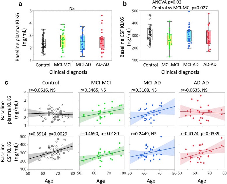
Fig. 4.

Kallikrein 6 (KLK6) levels among subject groups and association with age in cohort 2. One-way analysis is presented in box plots with individual subjects represented as dots. Plasma (a) and cerebrospinal fluid (CSF) (b) levels of KLK6 are compared between control subjects (white boxes), patients with mild cognitive impairment that remained unchanged over 24 months (MCI-MCI) (yellow boxes), patients with mild cognitive impairment who developed Alzheimer’s disease (MCI-AD) (blue boxes) and patients with Alzheimer’s disease (AD) (red boxes). c, d Linear correlation analysis between baseline plasma KLK6 (c) and baseline CSF KLK6 (d) values (y-axis) and age (x-axis). Black correlation lines = control subjects; green dots, green correlation lines = patients with MCI-MCI or patients with MCI stable over 24 months; blue dots, blue correlation lines = MCI-AD (MCI converters to AD = Clear dots); red dots, red correlation lines = AD from baseline. NS Not significant, ANOVA Analysis of variance
