
Fig. 3.
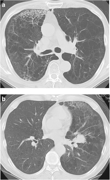
Computed tomography images of nivolumab-induced pneumonitis in Case 3. a Non-segmental ground-glass opacities and consolidations were observed in a predominantly subpleural distribution at both lungs on the 14th day of the initial nivolumab treatment. b After the predominant lesion at the right lung in the initial disease episode was reduced, the similar opacity was newly observed at the left lung in the time of relapse