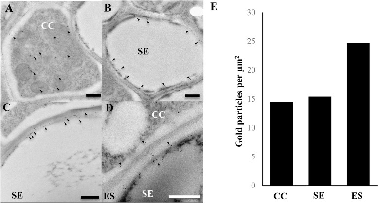
FIGURE 2.
Immunogold localisation of AtACBP3 using anti-AtACBP3 antibodies in transmission electron microscopy of cross sections of apical stems from 5-week-old Arabidopsis. (A) Companion cell. (B) Sieve element. (C,D) Extracellular space. (E) Densities of gold particles were quantified per μm2. Arrowheads indicate gold particles. Multiple floral stems from different individual plants sectioned and analysed in this experiment showed consistent results. Further images are available in Supplementary IEM Images. CC, companion cell; ES, extracellular space; SE, sieve element. Scale bar = 0.5 μm.

