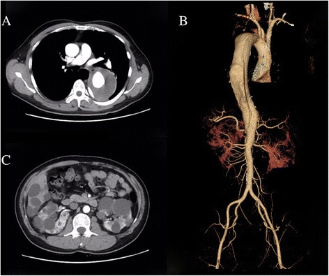
Fig. 2.

Radiographic findings of the proband. a Multi-slice computed tomography shows dissection aneurysm measuring 8.5 cm in diameter. b 3D–reconstructed computed tomography angiogram shows a Stanford B aortic dissection. c Multi-slice computed tomography shows multiple cysts in the liver and bilateral polycystic kidneys
