
Fig. 2.
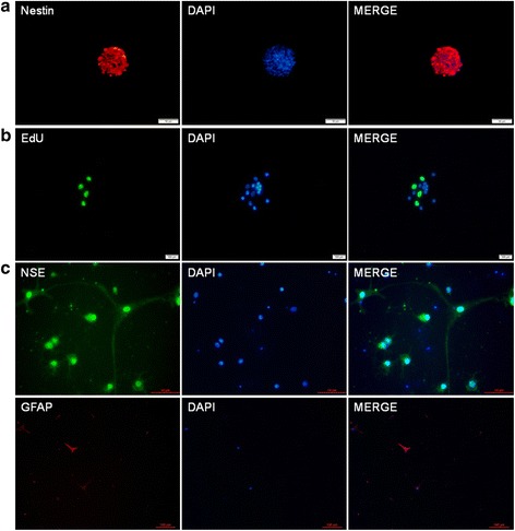
Immunofluorescent staining of NSCs derived from rat hippocampus. a Nestin immunoreactivity (red) was positive. b EdU immunoreactivity (green) was positive compared to control. c The immunofluorescence staining showed positive NSE (green) and GFAP (red) expression. DAPI (blue) was used to stain the nuclei