
Fig. 1.
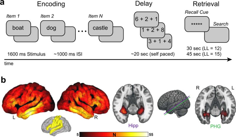
Recall task and electrode coverage. a. Experimental paradigm. On each trial, patients studied a list of words, performed a self paced arithmetic task, and finally recalled the items studied on the most recent list in any order. b. Electrode coverage. Left, the number of subjects with bipolar electrode centers within 10 mm of each vertex of the average cortical surface. Right, cross sections along the longitudinal axis of the MTL showing the number of subjects with bipolar electrode coverage localized within hippocampus (Hipp) or parahippocampal gyrus (PHG), extending 3 mm from electrode centroids. A priori anatomical regions of interest excluded from a subset of analyses are depicted in yellow. The depicted coverage discounts electrodes within the epileptogenic zone. L, left; R, right.