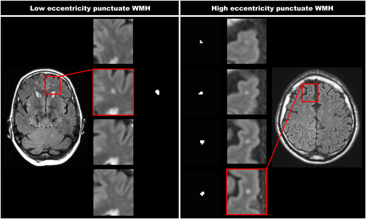
Figure 1.
Two WMH with a different eccentricity value. This figure represents two WMH that have a different eccentricity value. The shown FLAIR images have a voxel size of 0.96 × 0.96 × 3.00 mm3. The left panels show a punctuate deep WMH with a low eccentricity of 1.0 (close to spherical), which is seen in only one slice. The right panels show a punctuate deep WMH with a high eccentricity of 4.2 (strongly ellipsoidal), which is caused by the lesion extending in multiple slices. As can be appreciated, this difference in WMH shape can also be perceived visually.

