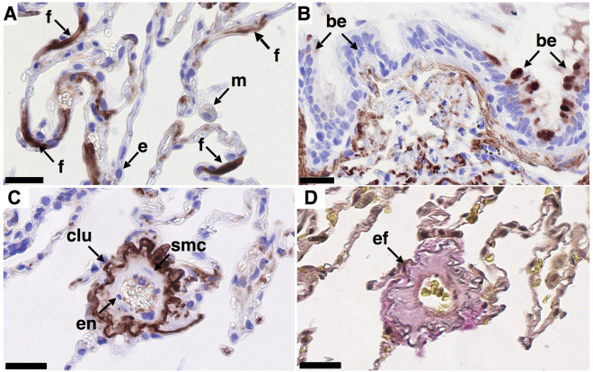
Figure 1.
Localization of clusterin in normal human lung. Clusterin was detected immunohistochemically by staining formalin-fixed, paraffin embedded 3 μm sections of human control lung tissue. Representative images of clusterin (clu, A-C, brown/red, nuclei - blue) and elastic fibers ((D), grey/black) in tissue obtained from control lung (n = 3). Clusterin localizes to fibroblast-like cells (A), to small areas of bronchial epithelial cells (B) and to elastic fibers in blood vessels and alveolar walls (C,D) serial sections). Clusterin was not detectable in macrophages, alveolar epithelial cells (A) or endothelial cells (C). Different cell populations/structures are indicated by arrows: f - fibroblast-like cell, m - macrophage, e – alveolar epithelial cell, be - bronchial epithelial cell, en - endothelial cell, smc – smooth muscle cell, ef - elastic fibers. Scale bar represents 25 µm.

