Figure 2.
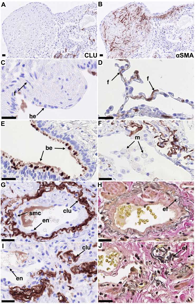
Localization of clusterin in IPF lung. Immunohistochemical staining for clusterin was performed on formalin-fixed, paraffin embedded 3 μm sections of IPF lung tissue (n = 3). Clusterin staining (clu, A, C-G, I brown/red, nuclei - blue) and staining for elastin (H, J, EvG, grey/black) in representative tissue sections. Clusterin is undetectable in αSMA positive myofibroblasts (A,B), forming and in cells overlying fibroblastic foci (A,C), compared to strong staining of fibroblast-like cells in morphologically normal non-fibrotic areas (D). Clusterin was observed sporadically in bronchial epithelial cells but more frequently than in controls (E). Similar to control lung, clusterin colocalized with elastin (G,H) and was undetectable in macrophages, smooth muscle and endothelial cells (F,G). Clusterin also colocalized with amorphous elastin aggregates in dense fibrotic regions (I,J). Different cell populations/structures are indicated by arrows; f - fibroblast-like cell, m - macrophage, he - hyperplastic epithelial cell, be - bronchial epithelial cell, en – endothelial cell, smc – smooth muscle cell, ef- elastic fibers. Scale bar represents 25 µm (A–J).

