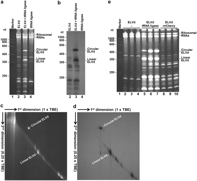
Figure 2.
Analysis of RNAs accumulated in E. coli cells that co-express ELVd RNA and eggplant tRNA ligase. RNAs were separated by PAGE, stained first with ethidium bromide (a,c and e), and transferred next to membranes, hybridized with a radioactive probe complementary to ELVd RNA and autoradiographed (b and d). (a and b) RNAs separated by single denaturing PAGE. Lane 1, RNA marker with the sizes of the standards indicated on the left in nt; lanes 2 to 4, RNAs from the E. coli cells transformed with pLELVd (lane 2), pLELVd and p15tRnlSm (lane 3) and p15tRnlSm (lane 4). (c and d) RNAs from the E. coli co-transformed with pLELVd and p15tRnlSm separated by two-dimension denaturing PAGE in two different buffer conditions, as indicated. (e) RNAs separated by single denaturing PAGE. Lane 1, RNA marker with the sizes of the standards indicated on the left in nt; lanes 2 to 10, RNAs from independent E. coli clones transformed with pLELVd (lanes 2 to 4), pLELVd and p15tRnlSm (lanes 5 to 7), and pLELVd and p15mCherry (lanes 8 to 10). In the different panels, the positions of the circular and linear ELVd RNAs, and of E. coli 23S and 16S rRNAs, are indicated. (a–d) E. coli cultures were grown at 25 °C and induced with 0.4 mM IPTG at OD600 0.6. Bacteria were harvested at 10 h post-induction. (e) E. coli cultures were grown at 37 °C and induced with 0.1 mM IPTG at OD600 0.1. Bacteria were harvested at 8 h post-induction. Each lane contains an aliquot of RNA that corresponds to 0.8 ml of culture in all cases.

