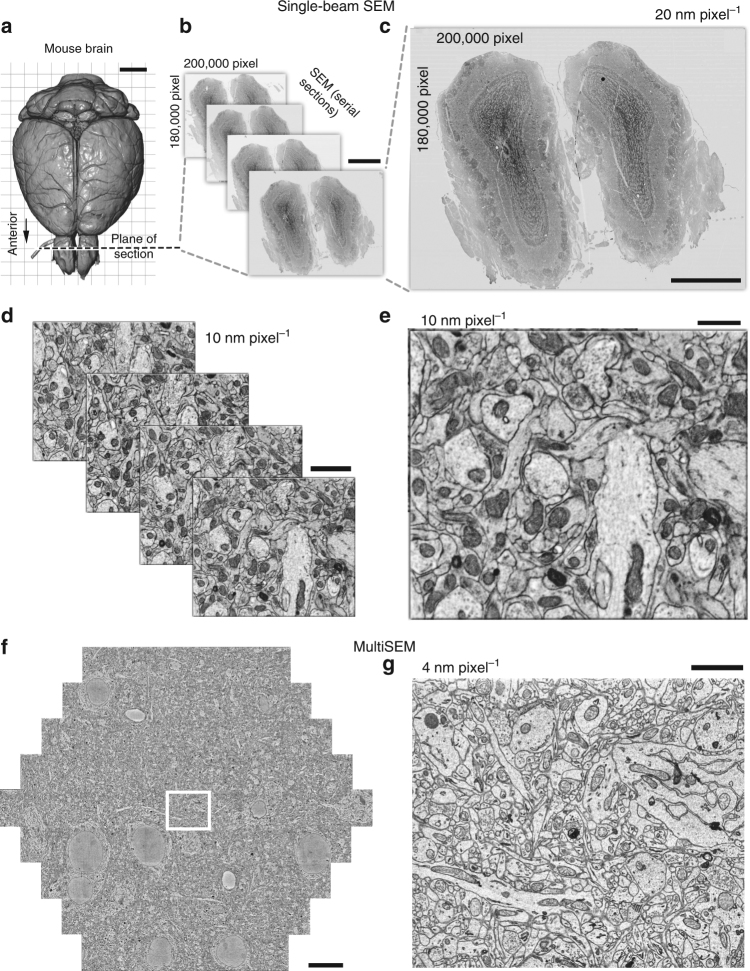
Fig. 7.
CNT tape provides good imaging conditions for large brain sections with single beam SEM (a-e) and MultiSEM (f, g). a 3D reconstruction of an adult mouse brain based on X-ray microCT. Scale, 2 mm. b Olfactory bulb serial sections (100 nm-thick) collected onto CNT tape are largely wrinkle-free and can be imaged with a BSE detector in high vacuum without charging. Scale, 1 mm. c Enlargement of an SEM mosaic, consisting of 3078 individual image tiles, with areas of the olfactory bulb indicated. Scale, 1 mm. d High-magnification stacks through the serial sections shows intact cellular membranes and well-stained synapses suitable for neuronal circuit reconstructions. Scale, 2 µm. e Enlarged serial section from d. Scale, 1 µm. f MultiSEM image of mouse primary somatosensory cortex captured with 4 nm pixel−1, 3128 × 2724 pixels image size for each beam, 100 ns dwell time and 1.5 keV landing energy. Stitched hexagon of ~35 nm thick section of cortical mouse brain tissue. Scale, 10 µm. g Enlarged single tile image outlined by the white rectangle in f. Scale, 2 µm

