Figure 3.
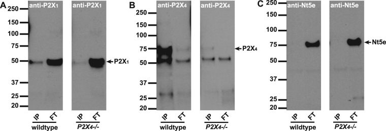
Immunoprecipitation with anti-P2X4 antibody. Antibodies to P2X4 were immobilized onto resin beads and then incubated with mouse bladder lysates to IP the antigen and co-IP interacting proteins. Proteins that were bound (IP: 2.5 µg protein/lane) or did not bind (FT: 25 µg protein/lane) to the beads, were resolved by SDS-PAGE, and Western blots were probed with A) P2X1, B) P2X4 or C) Nt5e antibodies. (A) Left and right panels show P2X1 immunoblots on IP and FT lysates from wild type and P2X4−/− mice. P2X1 is highly concentrated in the FT fractions. Minor potential P2X1 staining appears in the IP lane, however this is due to P2X4 antibody cross-reacting and pulling down some P2X1. (B) P2X4 antibody detects P2X4 as a band at 70 kDa in wild type IP lane, but is absent in P2X4−/− mice. The antibody shows minor cross-reactivity to P2X1 (50 kDa band). (C) An antibody to 5′-nucleotidase (Nt5e) demonstrates that pulldown with anti-P2X4 is ‘clean’ with no non-specific protein binding evident in the IP lanes.

