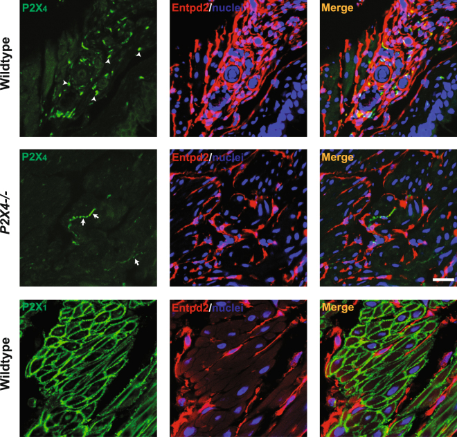
Figure 4.
P2X4 is expressed in bladder wall but not in BSM. Cryosections of mouse (wild type and P2X4−/−) bladders were labelled with antibodies to P2X4 (green), P2X1 (green), ENTPD2 (red), and Topro-3 to label nuclei (blue). Entpd2 (middle panels) labels interstitial cells among muscle bundles. Colour merged panels are shown on the right and merged signals are seen as yellow. White arrowheads (top left) indicate P2X4 labelling of unknown cellular structures. White arrows (middle left) indicate non-specific labelling by anti-P2X4 antibody in P2X4−/− mice. P2X1 receptors are abundantly expressed in BSM and that signal is clearly differentiated from interstitial cell staining (red). White scale bars = 10 µm.

