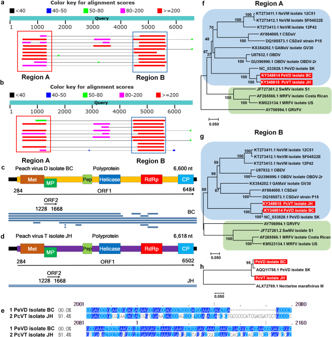
Figure 4.
Identification of a novel peach virus from two peach transcriptomes. (a) Visualization of BLAST result with the obtained PeVD isolate BC genome sequence. Red and blue boxes indicate region A and region B sequences, respectively, which show strong sequence similarity to other known viruses. (b) Visualization of BLAST results with the obtained PcVT isolate JH genome sequence. (c) Genome organization and the alignment of contigs associated with PeVD on the PeVD genome for the PeVD isolate BC. Two open reading frames (ORF)s: ORF1 and ORF2 are indicated by arrow lines with corresponding sequence positions. (d) Genome organization and the alignment of contigs associated with PcVT on the PcVT genome for PcVT isolate BC. (e) Comparison of nucleotide sequences between PeVD isolate BC and PcVT isolate JH. Phylogenetic trees of two PeVD isolates and a PcVT isolate and the associated known viruses based on regions A (f) and B (g) sequences. (h) Phylogenetic tree of two PeVD isolates and a PcVT isolate using polyprotein sequences. Nectarine marafivirus M was used as an outgroup.

