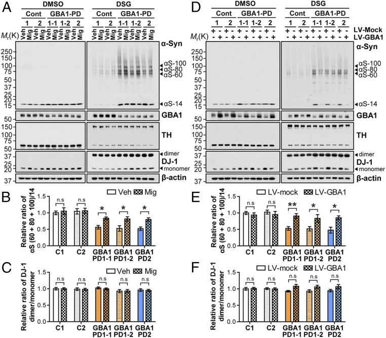
Fig. 4.
The formation of α-syn tetramers and related multimers in GBA1-PD iPSC-derived hDA neurons with miglustat treatment and lentiviral GBA1 transduction. (A) Cytosolic fractions of 1 mM DSG–cross-linked control and GBA1-PD hDA neurons with or without 100 μM miglustat treatment for 3 d were analyzed by Western blot using anti–α-Syn, anti-GBA1, anti-TH, anti–DJ-1, and anti–β-actin antibodies. (B) Quantification of the α-syn multimers to monomer ratio for A (n = 4). (C) Quantification of the ratio of DJ-1 dimers to monomer for A (n = 4). (D) Cytosolic fractions of 1 mM DSG–cross-linked control and GBA1-PD hDA neurons with LV-mock or LV-GBA1 viral transduction for 5 d were analyzed by Western blot analysis. (E) Quantification of the α-syn multimers to monomer ratio for D (n = 3). (F) Quantification of the ratio of DJ-1 dimers to monomer levels for D (n = 3). The error bars represent SEM. *P < 0.05; **P < 0.01; n.s., not significant.

