Fig. 3.
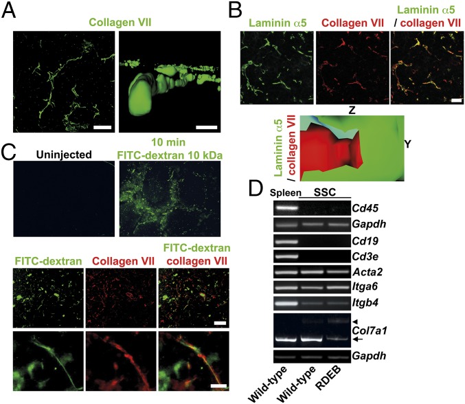
Collagen VII is part of lymphoid conduits. (A) Confocal analysis of splenic collagen VII structures (green). On the Left side is an XY projection, and the Right side shows a computed YZ 3D rendition. [Scale bars: 10 μm (Left); 1 μm (Right).] (B, Top) Maximum intensity projection of confocal Z stack of splenic follicle stained for laminin α5 (green) and collagen VII (red). (Scale bar, 10 μm.) (B, Bottom) A longitudinal cut, applied through computed 3D model of confocal Z-stack scans, shows collagen VII to be located inside the laminin α5-containing conduit basement membrane. The Y and Z axes are displayed as indicated. (C) Images of spleen sections from a wild-type mouse 10 min after systemic injection of 10-kDa FITC-dextran (green) stained for collagen VII (red). (Scale bars, 5 μm.) (D) RT-PCR analysis of mRNA from wild-type spleen, or cultured SSCs from wild-type or RDEB mice for Cd45, Cd19, Cd3e, Acta2, Itga6, Itgb4, Col7a1, and Gapdh transcripts. Absence of Cd45, Cd19, and Cd3e transcripts confirms cultures to be devoid of leukocytes and lymphocytes. Presence of Itgb4 transcript indicates follicular dendritic cells in the cultures (SI Appendix, Fig. S2) (31). Arrow points to wild-type Col7a1 transcript, and arrowhead to aberrantly spliced Col7a1 transcript (25).

