Fig. 2.
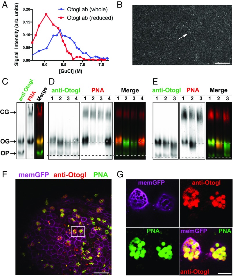
Otogl is a multimeric O-glycosylated glycoprotein. (A) Rate zonal centrifugation of nonreduced and reduced tadpole skin secretions probed with anti-Otogl antibody. (B) TEM of CsCl density gradient purified Otogl shows long-chain–like networks (arrow). (Scale bar: 200 nm.) (C) Coprobing of a Western blot of tadpole lysate with anti-Otogl antibody and PNA; a merged image is also shown. CG, cement gland mucin; OG, glycosylated form of Otogl; OP, precursor form of Otogl. (D) Treatment of tadpole lysate with O-glycosidase (2 h, lane 2, and 4 h, lane 3) and PNGase F (lane 4) compared with control (lane 1), probed with anti-Otogl and PNA; a merged image is also shown. Dashed lines represent the position of bands for control OG and OP. (E) Treatment of tadpole lysate with sialidase alone (lane 2) and sialidase + O-glycosidase (lane 3) compared with control (lane 1); a merged image is also shown. Dashed lines represent the position of bands for control OG and OP. In D and E, the signal for PNA from the cement gland mucin shows the approximately equivalent loading between lanes. (F) Image of section from fixed whole-mount tadpole skin with immunofluorescence for anti-Otogl and anti-GFP [labeling membrane-GFP (memGFP) to identify membranes] together with lectin histochemistry (PNA) shows colocalization of Otogl and PNA staining. The boxed area highlights two adjacent SSCs. (Scale bar: 50 μm.) (G) Zoomed-in image of boxed area from F shows Otogl and PNA colocalize within the vesicles of SSCs. ab, antibody. (Scale bar: 10 μm.)

