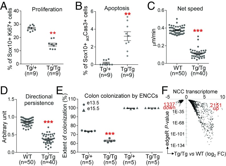
Fig. 2.
Global impairment of NCC development in ToupeeTg/Tg embryos. (A and B) Quantification of Ki67+ proliferating (A) and actCasp3+ apoptotic (B) NCCs (expressed in percentage of Sox10+ NCCs) in 30-μm transverse sections of E10.5 embryos at hindlimb level (ToupeeTg/+ vs. ToupeeTg/Tg). (C and D) Quantification of NCC migration speed (C) and movement persistence (D) in E10.5 embryos (WT;G4-RFP vs. ToupeeTg/Tg;G4-RFP). (E) Quantification of the extent of colon colonization by enteric NCCs (expressed in percentage of colon length from cecum to anus) at E13.5 and E15.5 (ToupeeTg/+;G4-RFP vs. ToupeeTg/Tg;G4-RFP). (F) Volcano plot summarizing a RNA-seq–based analysis of differential gene expression levels in E10.5 NCCs (WT;G4-RFP vs. ToupeeTg/Tg;G4-RFP). Only genes modulated at least 1.5-fold with a P value below 0.01 are displayed. **P ≤ 0.01 and ***P ≤ 0.001 (Student’s t test).

