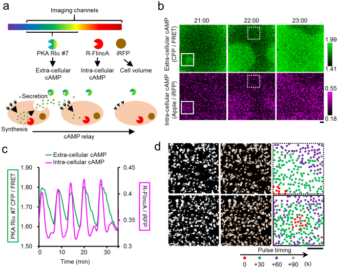
Figure 4.
Simultaneous imaging of cellular and environmental cAMP. (a) Intra-cellular synthesis and extra-cellular relay of cAMP in a population of D. discoideum cells, detected by R-FlincA and PKA RIα #7, respectively. Cytoplasmically expressed iRFP was the volume marker for correcting the motion artefact noise in R-FlincA signals. (b) The spatio-temporal dynamics of the environmental (top) and cellular (bottom) cAMP in a population of synchronously oscillating D. discoideum cells. Bar, 100 μm. (c) The population-averaged time courses of the [cAMP]in and [cAMP]ex in the solid inset in (b). (d) Pulse timing at the cell resolution in a wave receiving area (top, dashed inset in (b) and the centre of the de novo cAMP wave (bottom, solid inset in (b). Circles indicate the positions of individual cells. Colours discriminate the pulse timing. Bar, 100 μm. See also Supplementary Video S1.

