Fig. 3.
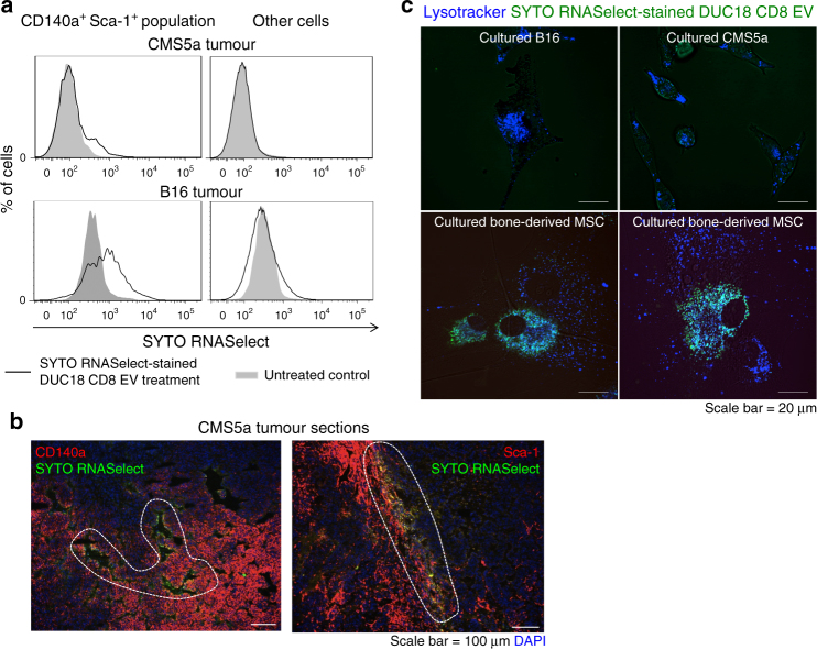
Rapid engulfment of EV RNAs by mesenchymal cells. a SYTO RNASelect-stained DUC18 CD8 EVs (10 μg [5–7 × 10⁸ vesicles]/tumour) were injected into CMS5a and B16 tumours (n = 3 per group). At 2 h after treatment, the cell suspensions from resected tumours were stained with PE-CD140a- and APC-Sca-1-specific mAbs, and analysed for SYTO RNASelect-derived green fluorescence in the CD140a+ Sca-1+ population or other cells, including tumour cells, by flow cytometry. b SYTO RNASelect-stained DUC18 CD8 EVs (10 μg [5–7 × 10⁸ vesicles]/tumour) were injected into CMS5a tumours (n = 3). At 2 h after treatment, the obtained tumours were sectioned and stained with PE-CD140a or PE-Sca-1-specific mAb. Each photo is a representative of 4–5 photos. The dotted circles indicate the overlapping areas of PE-originated red and SYTO RNSelect-derived green fluorescence. c SYTO RNASelect-stained DUC18 CD8 EVs (5 μg [4 × 10⁸ vesicles]/mL) were added to in vitro cultured B16, CMS5a or bone-derived MSCs (n = 5 per group). Two hours later, the cells were treated with lysotracker (blue), and then observed by confocal laser scanning microscopy. Each photo is a representative of images in 3–4 areas

