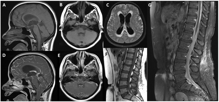
Figure 3.
Pre-contrast sagittal (a) and axial (b) T1-weighted images of the brain and spine (f) do not show any hyperintensity or T1 shortening. Post-gadolinium T1-weighted images (d–g) show diffuse leptomeningeal enhancement. Note the 7–8th cranial nerve enhancement (e). Axial fluid-attenuated inversion recovery image (c) reveals hyperintensity in the cerebral sulci and hydrocephalus.

